

How To Clean Armpit With Broken Clavicle
| Clavicle (collarbone) | |
|---|---|
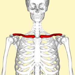 Collarbone (shown in carmine) | |
| Human collarbone | |
| Details | |
| Identifiers | |
| Latin | Clavicula |
| MeSH | D002968 |
| TA98 | A02.4.02.001 |
| TA2 | 1168 |
| FMA | 13321 |
| Anatomical terms of bone [edit on Wikidata] | |
The clavicle, or collarbone, is a slender, S-shaped apartment bone approximately half dozen inches (15 cm) long[1] that serves equally a strut betwixt the shoulder blade and the sternum (breastbone). There are two clavicles, one on the left and one on the right. The clavicle is the only long bone in the body that lies horizontally. Together with the shoulder blade, information technology makes upwards the shoulder girdle. It is a touchable bone, and in people who have less fat in this region, the location of the bone is clearly visible, as it creates a bulge in the skin. Information technology receives its name from the Latin clavicula ("little key"), considering the os rotates along its axis similar a key when the shoulder is abducted. The clavicle is the almost commonly fractured os. It can easily be fractured by impacts to the shoulder from the strength of falling on outstretched arms or by a straight hit.[2]
Construction [edit]
The collarbone is a thin doubly curved flat os that connects the arm to the body of the body. Located directly above the first rib, it acts as a strut to keep the scapula in identify so that the arm can hang freely. At its rounded medial cease (sternal stop), it articulates with the manubrium of the sternum (breastbone) at the sternoclavicular joint. At its flattened lateral end (acromial end), it articulates with the acromion, a process of the scapula (shoulder bract), at the acromioclavicular articulation.
The rounded medial region (sternal region) of the shaft has a long curve laterally and anteriorly along two-thirds of the entire shaft. The flattened lateral region (acromial region) of the shaft has an even larger posterior curve to articulate with the acromion of the scapula. The medial region is the longest clavicular region as it takes up 2-thirds of the entire shaft. The lateral region is both the widest clavicular region and thinnest clavicular region. The lateral end has a rough junior surface that bears a ridge, the trapezoid line, and a slight rounded project, the conoid tubercle (above the coracoid procedure). These surface features are attachment sites for muscles and ligaments of the shoulder.
It can be divided into three parts: medial end, lateral finish, and shaft.
Medial end [edit]
The medial cease is as well known equally the sternal end. It is quadrangular and articulates with the clavicular notch of the manubrium of the sternum to form the sternoclavicular joint. The articular surface extends to the inferior aspect for articulation with the first costal cartilage.
Lateral finish [edit]
The lateral cease is also known as the acromial end. It is apartment from above downward. It bears a facet that articulates with the shoulder to class the acromioclavicular joint. The area surrounding the joint gives an attachment to the joint capsule. The anterior border is concave forward and the posterior border is convex backward.
Shaft [edit]
The shaft is divided into two main regions, the medial region, and the lateral region. The medial region is also known as the sternal region, it is the longest clavicular region as it takes upward ii-thirds of the entire shaft. The lateral region is likewise known as the acromial region, it is both the widest clavicular region and thinnest clavicular region.

Lateral region of the shaft [edit]
The lateral region of the shaft has two borders and 2 surfaces.
- the anterior border is concave forward and gives origin to the deltoid muscle.
- the posterior border is convex and gives attachment to the trapezius muscle.
- the inferior surface has a ridge called the trapezoid line and a tubercle; the conoid tubercle for attachment with the trapezoid and the conoid ligament, office of the coracoclavicular ligament that serves to connect the collarbone with the coracoid process of the scapula.
Development [edit]
The collarbone is the outset os to brainstorm the process of ossification (laying down of minerals onto a preformed matrix) during evolution of the embryo, during the fifth and sixth weeks of gestation. However, information technology is i of the last basic to stop ossification at nearly 21–25 years of age. Its lateral end is formed past intramembranous ossification while medially it is formed by endochondral ossification. It consists of a mass of cancellous bone surrounded by a compact bone shell. The cancellous os forms via ii ossification centres, one medial and one lateral, which fuse later on on. The compact forms every bit the layer of fascia roofing the bone stimulate the ossification of adjacent tissue. The resulting meaty bone is known as a periosteal collar.
Even though information technology is classified as a long bone, the collarbone has no medullary cavity (marrow cavity) like other long bones, though this is not always true.[ citation needed ] It is fabricated up of spongy cancellous os with a shell of compact os.[3] It is a dermal bone derived from elements originally attached to the skull.
Variation [edit]
The shape of the clavicle varies more than most other long bones. It is occasionally pierced by a branch of the supraclavicular nerve. In males the clavicle is usually longer and larger than in females. A study measuring 748 males and 252 females saw a deviation in collarbone length between age groups eighteen–xx and 21–25 of well-nigh 6 and v mm (0.24 and 0.20 in) for males and females respectively.[four]
The left clavicle is commonly longer and weaker than the right clavicle.[3] [5]
The collarbones are sometimes partly or completely absent in cleidocranial dysostosis.
The levator claviculae musculus, present in 2–3% of people, originates on the transverse processes of the upper cervical vertebrae and is inserted in the lateral one-half of the clavicle.
Functions [edit]
The collarbone serves several functions:[iii]
- Information technology serves equally a rigid back up from which the scapula and free limb suspended; an arrangement that keeps the upper limb away from the thorax so that the arm has maximum range of motion. Acting as a flexible, crane-like strut, it allows the scapula to move freely on the thoracic wall.
- Roofing the cervicoaxillary canal, it protects the neurovascular parcel that supplies the upper limb.
- Transmits physical impacts from the upper limb to the centric skeleton.
Muscle [edit]
Muscles and ligaments that attach to the collarbone include:
| Zipper on collarbone | Muscle/Ligament | Other zipper |
|---|---|---|
| Superior surface and anterior border | Deltoid muscle | deltoid tubercle, anteriorly on the lateral third |
| Superior surface | Trapezius muscle | posteriorly on the lateral third |
| Inferior surface | Subclavius muscle | subclavian groove |
| Junior surface | Conoid ligament (the medial function of the coracoclavicular ligament) | conoid tubercle |
| Inferior surface | Trapezoid ligament (the lateral part of the coracoclavicular ligament) | trapezoid line |
| Inductive border | Pectoralis major muscle | medial third (rounded edge) |
| Posterior border | Sternocleidomastoid musculus (clavicular head) | superiorly, on the medial third |
| Posterior border | Sternohyoid muscle | inferiorly, on the medial third |
| Posterior edge | Trapezius musculus | lateral third |
Clinical significance [edit]
- Acromioclavicular dislocation ("AC Separation")
- Degeneration of the clavicle
- Osteolysis
- Sternoclavicular dislocations
A vertical line drawn from the mid-clavicle called the mid-clavicular line is used equally a reference in describing cardiac apex beat during medical exam. It is also useful for evaluating an enlarged liver, and for locating the gallbladder which is between the mid-clavicular line and the transpyloric plane.
Collarbone fracture [edit]
Clavicle fractures (colloquially, a broken collarbone) occur as a result of injury or trauma. The near common type of fractures occur when a person falls horizontally on the shoulder or with an outstretched hand. A directly hit to the collarbone will also cause a pause. In well-nigh cases, the directly hit occurs from the lateral side towards the medial side of the bone. The virtually common site of fracture is the junction between the two curvatures of the bone, which is the weakest point. This results in the sternocleidomastoid muscle lifting the medial aspect superiorly, which can consequence in perforation of the overlying pare.
Other animals [edit]
The clavicle starting time appears as role of the skeleton in primitive bony fish, where it is associated with the pectoral fin; they also accept a bone called the cleithrum. In such fish, the paired clavicles run behind and below the gills on each side, and are joined past a solid symphysis on the fish'south underside. They are, however, absent in cartilaginous fish and in the vast bulk of living bony fish, including all of the teleosts.[6]
The earliest tetrapods retained this arrangement, with the addition of a diamond-shaped interclavicle betwixt the base of the clavicles, although this is not found in living amphibians. The cleithrum disappeared early in the evolution of reptiles, and is not establish in any living amniotes, only the interclavicle is present in almost modernistic reptiles, and likewise in monotremes. In modernistic forms, all the same, in that location are a number of variations from the primitive pattern. For case, crocodilians and salamanders lack clavicles birthday (although crocodilians do retain the interclavicle), while in turtles, they form part of the armoured plastron.[vi]
The interclavicle is absent in marsupials and placental mammals. In many mammals, the clavicles are also reduced, or even absent, to allow the scapula greater liberty of motion, which may be useful in fast-running animals.[half-dozen]
Though a number of fossil hominin (humans and chimpanzees) clavicles take been found, well-nigh of these are mere segments offering express data on the form and function of the pectoral girdle. One exception is the clavicle of AL 333x6/nine attributed to Australopithecus afarensis which has a well-preserved sternal end. One interpretation of this specimen, based on the orientation of its lateral stop and the position of the deltoid zipper area, suggests that this clavicle is distinct from those constitute in extant apes (including humans), and thus that the shape of the man shoulder dates dorsum to less than 3 to 4 million years ago. Nevertheless, analyses of the clavicle in extant primates suggest that the depression position of the scapula in humans is reflected mostly in the curvature of the medial portion of the clavicle rather than the lateral portion. This part of the bone is similar in A. afarensis and it is thus possible that this species had a loftier shoulder position like to that in modern humans.[seven]
In dinosaurs [edit]
In dinosaurs the main basic of the pectoral girdle were the scapula (shoulder bract) and the coracoid, both of which directly articulated with the clavicle. The clavicle was present in saurischian dinosaurs merely largely absent in ornithischian dinosaurs. The identify on the scapula where information technology articulated with the humerus (upper bone of the forelimb) is the chosen the glenoid. The clavicles fused in some theropod dinosaurs to course a furcula, which is the equivalent to a wishbone.[viii]
In birds, the clavicles and interclavicle take fused to class a unmarried Y-shaped bone, the furcula or "wishbone" which evolved from the clavicles found in coelurosaurian theropods.
Additional media [edit]
-

Position of collarbone (shown in reddish). Blitheness.
-

Shape of collarbone (left). Animation.
-
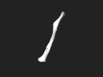
3D epitome
-

Pectoral girdle—front
-

Diagram of the human shoulder joint, forepart view
-
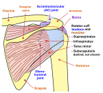
Diagram of the human shoulder joint, dorsum view
-

Muscles of the neck. Inductive view.
-

Clavicle
Come across besides [edit]
- Clavicle fracture
References [edit]
- ^ The Shoulder Complex. In: Prentice We. eds. Principles of Athletic Grooming: A Guide to Evidence-Based Clinical Practice, 17e. McGraw-Hill; Accessed Oct 30, 2022. https://accessphysiotherapy-mhmedical-com.libaccess.lib.mcmaster.ca/content.aspx?bookid=2992§ionid=250962289
- ^ "Busy Bones". 2022-05-xiii. Retrieved 2016-12-02 .
- ^ a b c Moore, Keith L.; Dalley, Arthur F. (1999). Clinically Oriented Anatomy (4th ed.). Lippincott Williams & Wilkins. ISBN978-0-683-06141-3.
- ^ "medind.nic.in" (PDF). Archived (PDF) from the original on 2022-03-27. Retrieved 2012-02-thirteen .
- ^ A. Bernat, T. Huysmans, F. Van Glabbeek, J. Sijbers, J. Gielen, and A. Van Tongel (2014). "The beefcake of the clavicle: A Three-dimensional Cadaveric Study". Clinical Beefcake. 27 (five): 712–723. doi:ten.1002/ca.22288. PMID 24142486. S2CID 23982787.
{{cite journal}}: CS1 maint: multiple names: authors list (link) - ^ a b c Romer, Alfred Sherwood; Parsons, Thomas Southward. (1977). The Vertebrate Body. Philadelphia, PA: Holt-Saunders International. pp. 184–186. ISBN978-0-03-910284-v.
- ^ Larson, Susan G. (2009). "Evolution of the Hominin Shoulder: Early on Homo". In Grine, Frederick Eastward.; Fleagle, John G.; Leakey, Richard Due east. (eds.). The First Humans - Origin and Early Evolution of the Genus Human being . Vertebrate Paleobiology and Paleoanthropology. Springer. p. 66. doi:x.1007/978-one-4020-9980-9. ISBN978-ane-4020-9979-3.
- ^ Martin, A.J. (2006). Introduction to the Study of Dinosaurs. Second Edition. Oxford, Blackwell Publishing. pg. 299-300. ISBN 1-4051-3413-5.
External links [edit]
- Clavicle [ permanent dead link ] - BlueLink Anatomy - University of Michigan Medical School
| | Wikimedia Commons has media related to Clavicle. |

Source: https://en.wikipedia.org/wiki/Clavicle
Posted by: villagomezwoperand.blogspot.com
0 Response to "How To Clean Armpit With Broken Clavicle"
Post a Comment